Microscope and imaging
Root canals can be narrower than a pencil lead. Magnification and good images are how an endodontist sees them.
Mountain View 650-691-0999Saratoga 408-252-5700
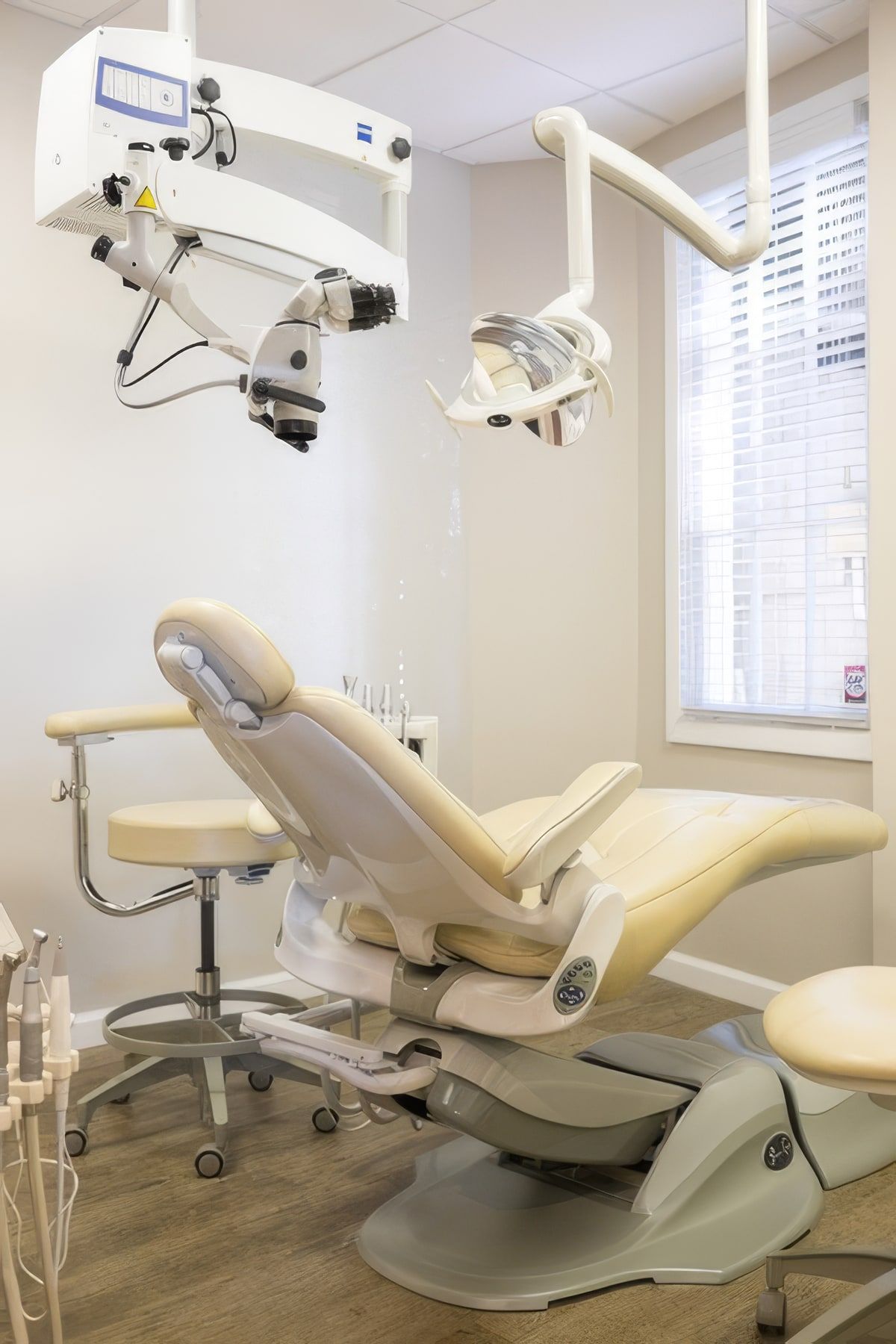
The surgical microscope
The microscope sits above the chair and magnifies the inside of the tooth, with light that shines straight down into it. It helps Dr. Hirose find small extra canals, see fine cracks, and clean and seal each canal precisely.
It makes a measurable difference. In a clinical study of 312 upper molars treated by endodontists, those using magnification found the hidden second canal about three times as often as those using none. Teeth with a missed canal are 4.4 times more likely to develop infection at the root tip, according to a study of 1,397 3D scans from the University of Pennsylvania.
Microsurgery
When a root tip needs treatment, the microscope matters even more. A meta-analysis of the research found success rates of 94 percent for modern endodontic microsurgery and 59 percent for older techniques. Read about apicoectomy and microsurgery.
X-rays
X-rays show the roots, the bone around them and any sign of infection. Dr. Hirose takes the images your tooth needs and shares them, with a report, with your dentist.
- Buhrley LJ et al. Effect of magnification on locating the MB2 canal in maxillary molars. J Endod, 2002.
- Karabucak B et al. Prevalence of apical periodontitis in endodontically treated premolars and molars with untreated canal. J Endod, 2016.
- Setzer FC et al. Outcome of endodontic surgery, a meta-analysis of the literature, part 1. J Endod, 2010.
Relief starts with a call
Call the office closest to you. If you are in pain, say so when you call, and the team will look for the soonest opening at either office.
Request an appointment-
Mountain View
100 West El Camino Real, Unit 67
-
Saratoga
18988 Cox Avenue, Suite D